PHILADELPHIA – The normal functioning of our hearts is maintained by our body’s control center - the brain - via an intricate network of nerves. When this communication is disrupted, it results in heart disease, including heart attacks, sudden cardiac death and problems in blood supply. As an added layer of safety, the heart has its own ‘little brain’, called the intracardiac nervous system (ICN) to monitor and correct any local disturbances in communication. The ICN is essential in supporting heart health and can even protect cardiac muscle during a heart attack. But it’s not clear how exactly the ICN carries out these roles, because the organization of the neurons that make up the ICN are poorly understood; we don’t know where they are located in the heart, how they are connected to each other, and what their molecular properties are.
In a groundbreaking study published in iScience on May 26th, researchers at Thomas Jefferson University and their collaborators have been able to answer these questions in unprecedented detail.
“The ICN represents a big void in our understanding that falls between neurology and cardiology,” says co-senior author James Schwaber, PhD, director of the Daniel Baugh Institute for Functional Genomics and Computational Biology (DBI) and co-senior author of the study. “Our goal was to bridge that gap by providing an anatomical framework of the ICN and a foundation to understand its role in heart health.”
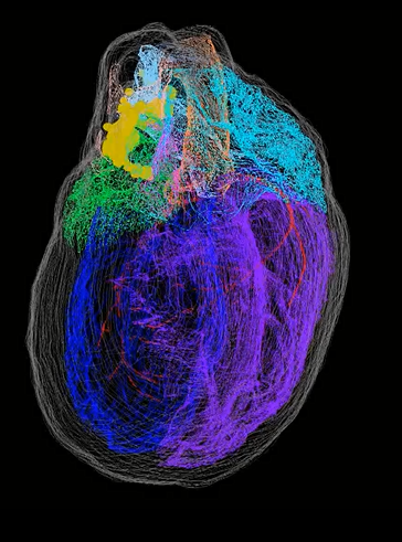
“The only other organ for which such a detailed high-resolution 3D map exists is the brain,” says co-senior author Raj Vadigepalli, PhD, Professor of Pathology, Cell Biology and Anatomy. “In effect what we have created is the first comprehensive roadmap of the heart’s nervous system that can be referenced by other researchers for a range of questions about the function, physiology, and connectivity of different neurons in the ICN.”
The study drew on technologies and expertise from different research groups (from Jefferson and University of Central Florida) and industry partners (Strateos and MBF Bioscience), eventually creating a dual-approach pipeline. One approach involved a novel imaging technique called Knife-Edge Scanning Microscopy (KSEM) that allowed the researchers to build a precise 3D model of the entire rodent heart; it is the first use of this technology for cardiac research. The second approach used a technique called laser capture microdissection to sample single neurons for gene expression analysis, as well as to precisely map their individual positions within the 3D structure of the heart.
“Because this hasn’t been done before, we were trouble-shooting the protocol as we went along,” says co-author Sirisha Achanta, Lab Manager at the DBI. “The heart, unlike the brain, is not symmetrical, so we had to figure out ways to maintain consistency across each heart that we imaged.”